Breadcrumbs
- Home
- About
- LMP Art Competition
- 2021 submissions & winners
LMP Art Competition winners 2021
In June 2021, we asked the LMP community to show us the beauty of their slides to share our love of the images that we see under the microscope.
Our students, learners, faculty, staff, and alumni sent in many beautiful images and it was a difficult task for the LMP leadership and administrative staff to vote on winners.
The top three images receive a cash prize, will have their image featured on our strategic plan, and will be framed for display in the Medical Sciences Building.
Enjoy the winners, and all submissions, of our art competition!
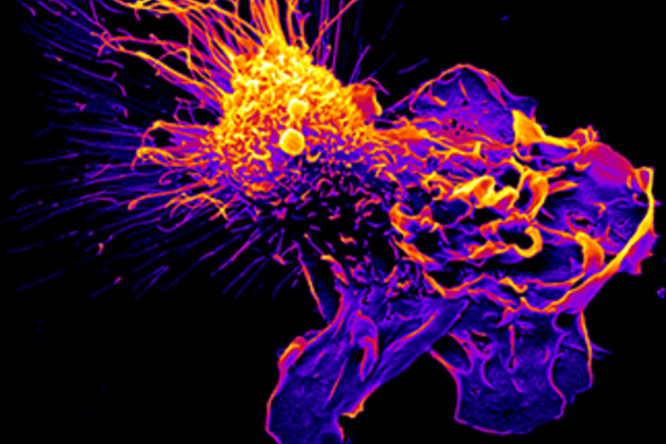
First place: 'Mac Attack'
Daniel Schlam, Resident, Anatomical Pathology postgraduate program
About Daniel's image
A scanning electron micrograph of a human macrophage fighting a fungal infection. Having sensed danger (in the form of soluble anaphylatoxins), this macrophage extends dramatic actin-driven extensions and membrane ruffles, which allow for tracking, trapping, and eventually internalizing, the invading pathogens.
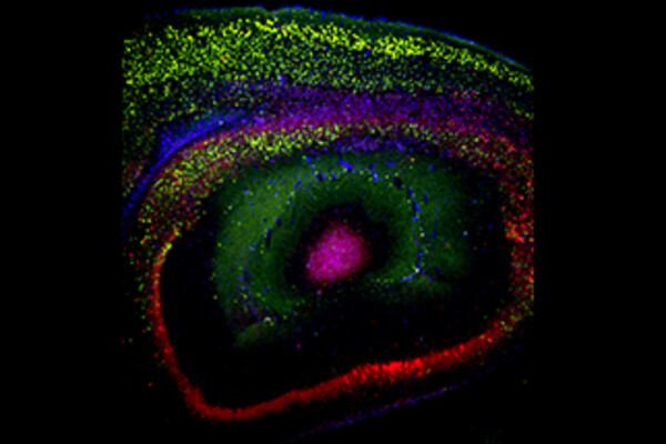
Second place: untitled
Lilian Lin, PhD Candidate in Dr. Janice Robertson's lab
About Lilian's image
An immunofluorescence image of a mouse's brain that has been injected with the adeno associated virus (AAV) that expresses EGFP tagged TDP-35, a pathological splice variant of TDP-43. TDP-43 pathology is seen in ALS.
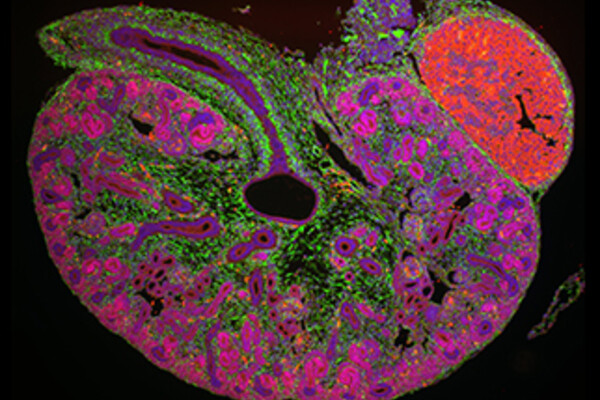
Third place: 'Murine Embryonic Kidney'
Robert D'Cruz, MD/PhD Candidate in Dr. Norman Rosenblum's lab
About Robert's image
A midsagittal section of an E15.5 murine embryonic kidney imaged using a scanning confocal microscope. The embryonic kidney has been immunolabelled with DAPI to label nuclei (blue), SALL1 to label nephron and stromal progenitor cells (red), and VIMENTIN to label interstitial cells (green).
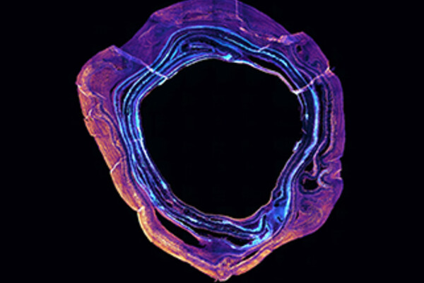
An honourable recognition for 'Ring of fire'
Aaryn Montgomery-Song, MSc student in Dr. Rita Kandel's lab
There were so many beautiful images that it was hard just to pick a top three. Aaryn's image came in so close that we felt we should give it an 'honourable recognition' and include it as one of the images to be framed in MSB.
About Aaryn's image
A tissue-engineered intervertebral disc stained for type I and II collagens visualized by epifluorescence microscopy. The inner rings of the disc are unique for their expression of type II collagen, whereas the outer rings only express type I collagen. In this image, miniature lab-grown discs follow a similar distribution of collagens. Type I collagen pseudo is coloured magenta/yellow and type II pseudo coloured cyan/white.
All submissions
It was a very difficult decision to choose a winner which you will see from all the wonderful entries below. Thank you to all who submitted an image.
Enjoy the beauty of LMP slides!
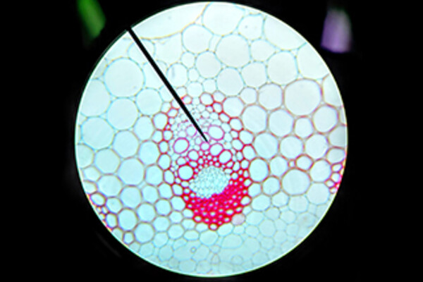
Kai Yuan Chen, Undergraduate Student
The xylem and phloem of a plant's vascular tissue under a light microscope.
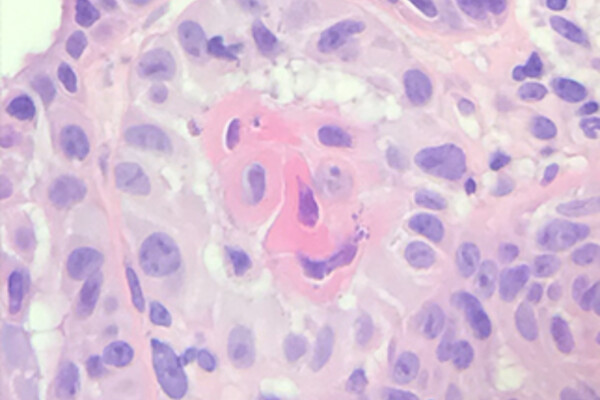
Valerie Dube, LMP faculty
'Squamous Monster': Squamous cell carcinoma, with an optical microscope.
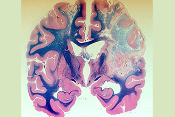
Juan Bilbao, LMP faculty
'Brain infarcts secondary to emboli': Whole brain embedding in celloidin, sledge microtome section. Staining: Luxol-Fast-Blue, Haematoxylin-Eosin.
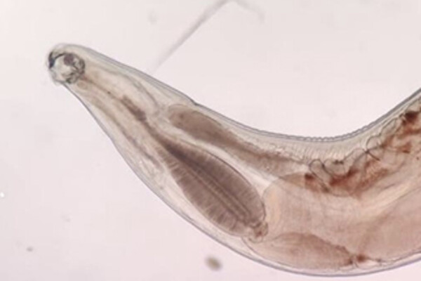
Ruchika Gupta, Postgraduate Trainee
'Beauty of the beast': Female of Anyclostoma duodenale. Light microscopy, wet mount picture from gastric aspirate in a patient with severe gastritis and GI bleeding.
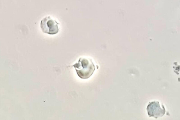
Melika Loriamini, Graduate Student
'MMA (mononuclear monolayer assay)': Opsonized RBCs phagocytes by monocyte from PBMCs of healthy donor, under Phase contrasts microscope.
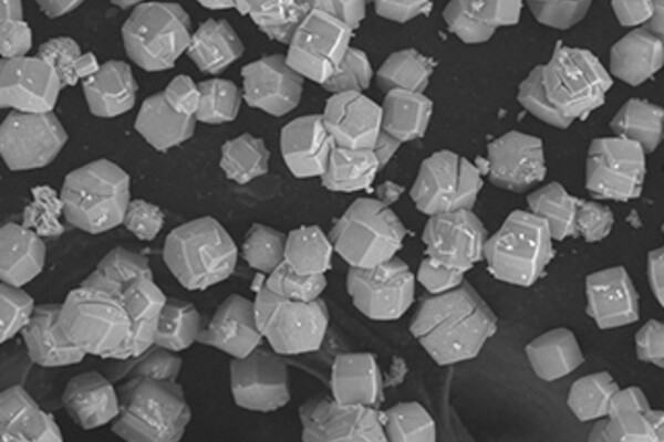
Vida Maksimoska, Graduate Student on the Translational Research Program
'Salts of Science': Using Scanning Electron Microscopy, salts synthesized for being used in wound dressings. The synthesis process produces quite uniform cuboidal crystals.
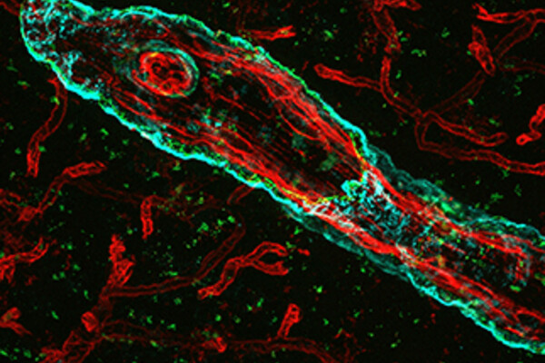
Mingzhe Liu, Graduate Student
'Cerebral Amyloid Angiopathy - Not Just a Plaque Story': A 40x immunofluorescent image taken of a coronal TgF344-AD (transgenic Alzheimer's disease) rat brain section. This tissue was stained for 4G8 (anti Amyloid-beta antibody), α-SMA (arterioles), and lectin (blood vessels). In the foreground, we can see a build up of cerebral amyloid angiopathy (CAA) on the arteriole. And in the background, we can see the rest of the diverse cerebrovascular system.
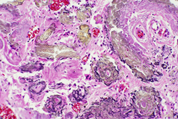
Rachel Jug, Postgraduate Trainee
'Pathologic Treasures': Image of Gamna-Gandy bodies from the spleen of a patient with sickle cell disease who died following sickle cell crisis leading to bone marrow infarction that embolized, passed through patent atrial septal defect, and obstructed a coronary artery. Light microscopy image, stained with hematoxylin and eosin, 20x magnification.
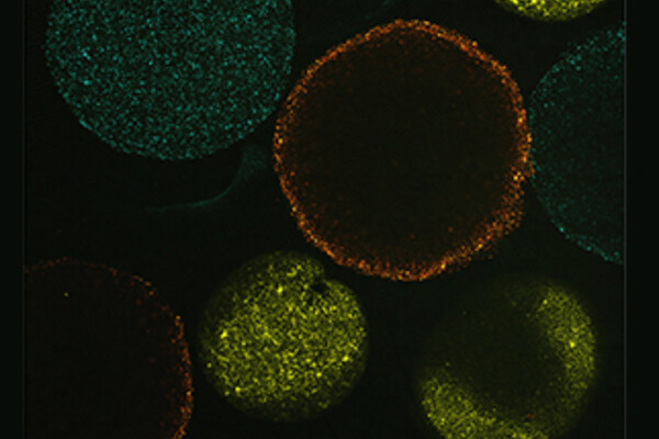
Joshua Rosen, Graduate Student
These are fibroblasts we derived from patients with non-small cell lung cancer. The cells were cultured in hydrogel to assess contractility, and were stained with different plasma membrane dyes. We used widefield fluorescence microscopy (with tiling function) to obtain this image.
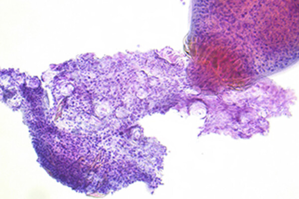
Heather Ruff, LMP faculty
'Mourning the dead': A Pap-stained ThinPrep slide from a renal cyst that shows Echinococcus. In this image an Echinococcus scolex is seen next to the fragmented remains of an adult Echinoccus cestode, with the calcareous corpuscles clearly visable in the fragmented adult.
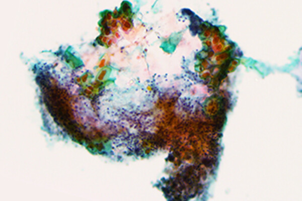
Chiyun (Angela) Wang, Postgraduate Trainee
'Sea Turtle's Migration': Papanicolaou-stained ThinPrep cytology of urine from ileal conduit showing vegetable cells originating from the ostomy adhesive. They reminded me of the pattern on a turtle's shell and how sea turtles make use of currents during their long-distance migrations.
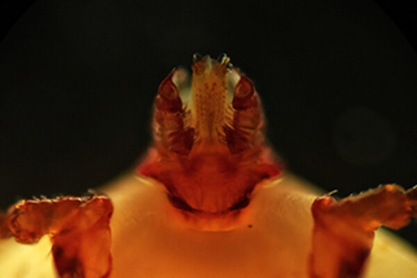
Fabio Tironi, Postgraduate Trainee
'Hitchhiker': A living tick on the slide. Zeiss Axio Lab A1 (optical microscope, Moto Z2 Play cell phone camera over ocular lens). A homeless dog entered our Forensic Institute (in Southern Brazil) by the backdoor. Skinny, covered with ticks, he was fed by our team, taken to a vet and forwarded to a lovely home. Some ticks remained as visitors for a couple of days...
Bojana Djordjevic, LMP faculty
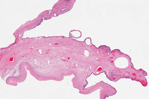
'Ovarian mucinous cystadenoma - Lab Rat': HE image. Ovarian endometriosis. This is not a sad rat that is trapped in the lab. No. This lab rat has made the lab its home. It knows every nook and cranny of every space. It delights in traveling its secret corridors after hours. It loves carrying out experiments, looking at slides and discussing cases with its lab rat friends. Sure, it might have grown a few warts on its back over the years, and become a bit sensitive to sunlight, but these are badges of honor, of life happily lived, of time well spent!
Andrew Wang, Alumnus from our MSc program
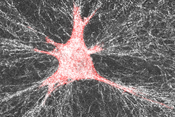
'A Spider on its Web': While this image may look like a spider on its web, it is actually an NIH 3T3 fibroblast on a collagen scaffold! This fibroblast was modified to overexpress a protein called DDR1, which is upregulated in fibrosis and certain cancers. We have previously shown that DDR1-overexpressing cells pull on collagen fibers as they spread, to increase the number of aligned (parallel) fibers, and collagen stiffness, which are important hallmarks of fibrotic disease which can be seen in this image. Images were obtained through confocal reflectance microscopy, where the red staining represents F-actin and white represents individual collagen fibers.
