Breadcrumbs
- Home
- About
- LMP Art Competition
- 2022 submissions & winners
LMP Art Competition winners 2022
Every spring, we ask the LMP community to show us the beauty of their slides to share our love of the images that we see under the microscope.
Our students, learners, faculty, staff, and alumni sent in many beautiful images and all members of the LMP community could vote for their favourites.
The top three images receive a cash prize, will receive canvas of their image, and it will be framed for display in the Medical Sciences Building.
Enjoy the winners, and all submissions, of our art competition!
First place: 'Lights, Camera, Astroglia in Action!'

Ain Kim, MSc candidate in Dr. Gabor Kovac's lab
About Ain's image
An immunofluorescence image of a mouse primary astroglia isolated from adolescent mouse brain tissue using GLAST magnetic bead cell separation. This astroglia flaunts its beauty under the widefield fluorescent microscope while we study their role in many neurodegenerative diseases such as Alzheimer’s and Parkinson’s Disease.
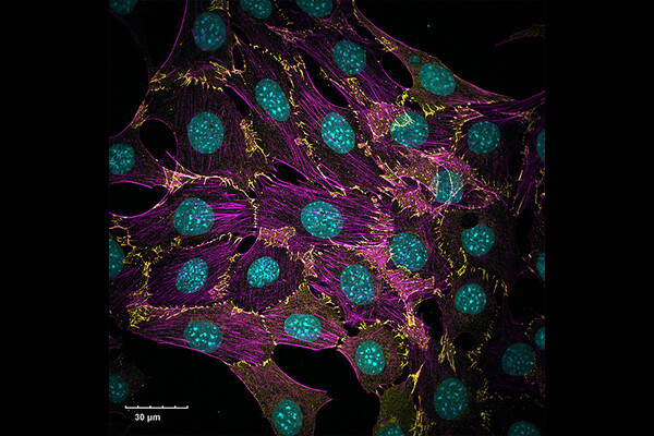
Second place: 'Cellular Connections'
Jonah Burke-Kleinman, PhD candidate in Dr. Michelle Bendeck's lab
About Jonah's image
N-Cadherin (yellow) junctions at cell-cell contacts between vascular smooth muscle cells, also stained for filamentous actin (magenta) and cell nuclei (cyan). These cells were plated on glass coverslips and imaged with an Olympus FV3000 laser-scanning confocal microscope using a 60x objective lens.
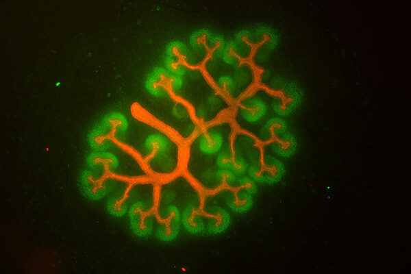
Third place: 'Branching Kidney'
Robert D'Cruz, MD/PhD Candidate in Dr. Norman Rosenblum's lab
About Robert's image
E12.5 murine embryonic kidney explant cultured for 72 hours and then immunofluorescently stained for CYTOKERATIN (red) to mark the epithelial branching network and SIX2 (green) to label the immature developing nephrons. Kidney explant was imaged using a spinning disk confocal microscope.
All submissions
It was a very difficult decision to choose a winner which you will see from all the wonderful entries below. Thank you to all who submitted an image.
Enjoy the beauty of LMP slides!
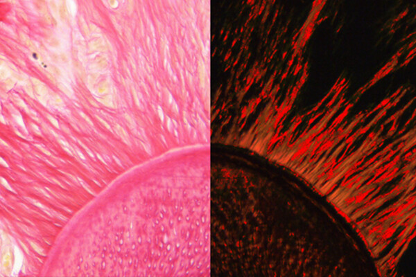
Aiman Ali, Research staff
'The Sun under our microscope': Horizontal cut of mouse tooth. H&E stain / Picrosirius red stain. Optical microscope X10.
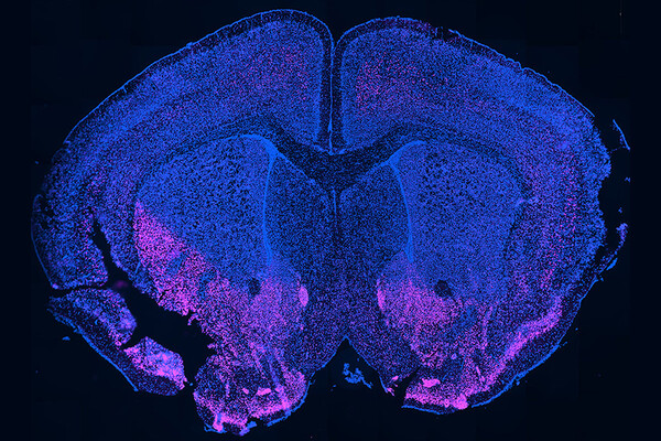
Shawar Ali, graduate student
'To Bcl11b, or not to Bcl11b, that is the question': A fluorescence microscopy image of a wild type adult mouse brain coronal section. Immunostaining was performed using Hoechst dye (blue) to stain nuclei and rabbit anti-Bcl11b antibody (magenta) to stain the neuronal transcription factor Bcl11b. Here, we were testing a new rabbit anti-Bcl11b antibody to see if was suitable for our experiments, hence the title of the image.
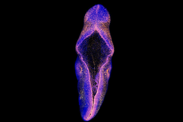
Polina Balin, Graduate student
'untitled': "This is a whole mount staining of E9.5 murine embryo with a mesenchymal and dorsal neural tube marker Msx1 in orange and nuclear marker DAPI in blue. Confocal microscope Nikon A1R was used to scan the embryo and build the z-stack. The micrograph depict maximum intensity projection of the signal. The micrograph shows dorsal (back) view of the embryo.
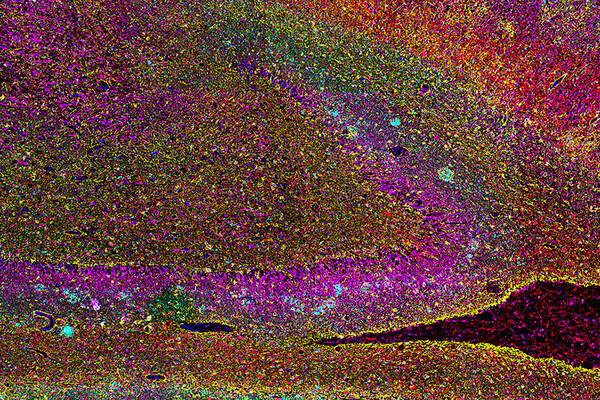
Seojin Lee, Graduate student
'The Colours of Memory': Hippocampus of Alzheimer's Disease human brain stained with six different immunofluorescence markers, visualizing neurons and astrocytes with iron homeostatic and pathologic-hallmark proteins.
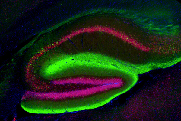
Lilian Lin, Graduate Student
'In Your Mind's Eye': This is a fluorescence microscopy image of the hippocampus of a mouse brain. The brain is stained with nuclear staining inn blue, neuronal staining in red, and green is a pathological form of an ALS-associated protein called TDP-35.
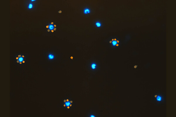
Melika Loriamini, Graduate Student
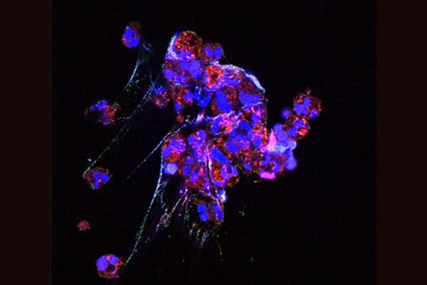
'Rosette Formation': Rosette formation by PBMCs and opsonized RBCs with anti-D (WinRho) under a fluorescent microscope-Monocytes stained with Hoescht are indicated in blue, while opsonized RBCs stained with PKKH are indicated in red. At room temperature, opsonized RBCs adhere to the FcγR on monocyte membrane and form a rosette, as determined by MMA (monocyte-mononuclear assay).
Maria Maqsood, Graduate Student
'Complement mediated neutrophil activation via the process of NETosis': In this image we used Immunofluorescence confocal imaging to examine neutrophils (a type of white blood cells) that were stimulated via complement by first presensitizing the cells with anti-CD59 antibody and then exposing them to normal human serum which contains all the necessary complement proteins to initiate both the classical and alternative pathway directly on their surface.
Tyler Redublo, Graduate Student
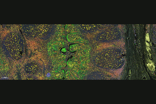
'Tropical Tonsil': Human tonsil tissue stained using multiplex immunohistochemistry, in which fluorescent dyes are used to detect protein localization. 6 different biomarkers, as well as DAPI counterstain, were detected simultaneously within a single image, using a multi-spectral microscope.
Mitra Shokrollahi, Graduate Student
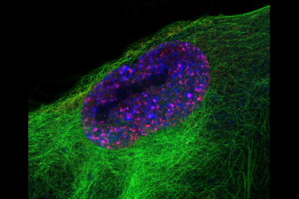
'Early cellular response to DNA damage': A fibroblast isolated from the normal lung tissue (IMR-90 cell) visualized by immunofluorescence and Airyscan confocal microscopy after induction of DNA damage. Blue shows DNA in the nucleus, green shows microtubules and red shows phosphorylated histone variant H2AX which constitutes about 2-25% of the H2A histones in mammalian chromatin. H2AX becomes phosphorylated, then called γH2AX, as an early reaction to DNA double-strand breaks. γH2AX generally reflects the presence of double-strand breaks in DNA.
Gino Somers, LMP faculty
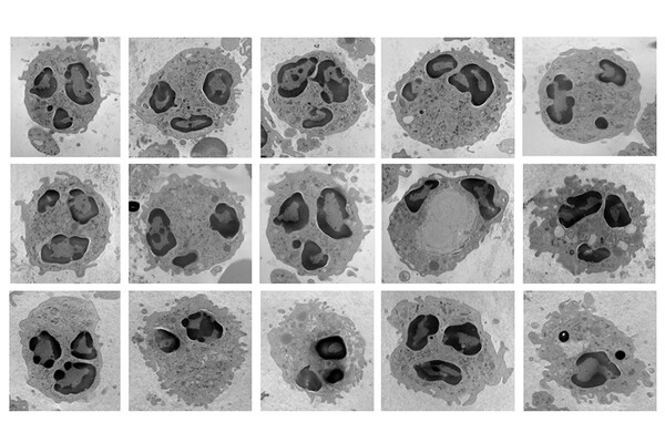
'Neutrophil emojis': Transmission electron microscopy of neutrophils at 12,000 times magnification.
Yuetong Song, Graduate Student
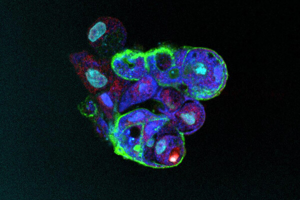
'3D Budding': This is an image of human induced pluripotent stem cell derived 3D lung airway organoid. Green is EPCAM, Red is SCGB3A2, Dark blue is KRT5 and Baby blue is DAPI. I used the leica confocol microscope and it is under 40X magnification. IPSC need 1 month to direct-differentiatie into lung epithelial progenitor cells. Then, these organoids are generated by lung epithelial progenitor cells sorting. Before imaging this 3D airway organoid, I need to stain them under disectional microscopy using p200 pipette since they are very tiny.
Manal Tadros, LMP Faculty
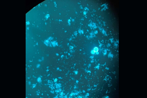
'A Blast-O-Fluorescence': Blastomyces dermatitidis (dimorphic fungus) in its yeast state in a respiratory sample. Under the fluorescent microscope with its characteristic broad base budding.
Qianghua Zhou, Postgraduate Trainee
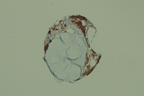
'Nautilus': Optic microscope, IHC staining of a bone marrow biopsy specimen.
