Breadcrumbs
- Home
- About
- LMP Art Competition
- 2023 submissions & winners
LMP Art Competition winners 2023
Every spring, we ask the LMP community to show us the beauty of their slides in the LMP Art Competition to share our love of the images that we see under the microscope.
Our students, learners, faculty, and staff sent in many beautiful images and all members of the LMP community were invited to vote for their favourites online.
The top three images receive a cash prize and a canvas of their image which will be displayed at the Annual Celebration of Excellence on June 22, 2023.
Enjoy the winners, and all submissions, of our art competition!
See submissions and winners from:
Honourable Recognition
This year, we give a special, honourable recognition to two fantastic images that really stood out.
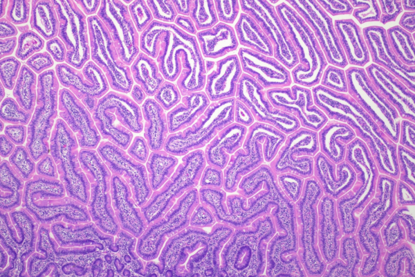
The Great Intestinal Reef
Kevin Kuan, Gastrointestinal Pathology Fellow
About Kevin's image
A light microscopy image of intestinal villi from the jejunum. Hematoxylin & eosin stained.
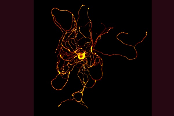
Reaching out in the darkness
Shane Eaton, Neuropathology Resident
About Shane's image
Isolated sensory neurons reach out with innumerable processes, searching for their target. Immunofluorescence image of a murine dorsal root ganglion sensory neuron in primary cell culture. Immunostained for neurofilament and βIII-tubulin.
Prize winners
The following received the top votes from the LMP Community.
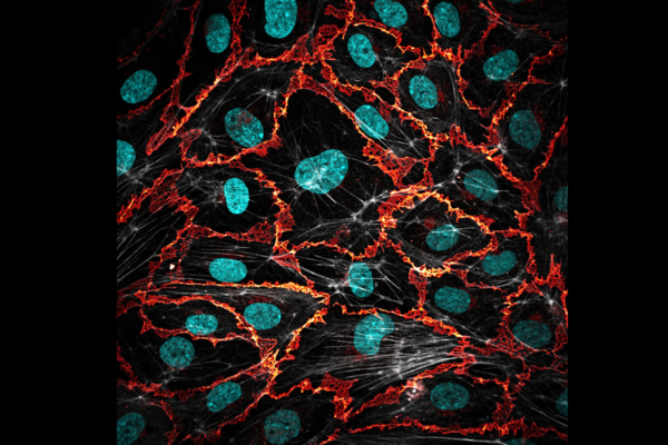
First place 'A Permeable Barrier'
Jonah Burke-Kleinman, PhD candidate in Dr. Michelle Bendeck's lab
About Jonah's image
Endothelial cells line all the blood vessels of our bodies and form a semipermeable, thrombo-resistant container for the blood. This image was obtained by laser-scanner confocal immunofluorescence microscopy of human umbilical vein endothelial cells (HUVECs) cultured in vitro. Cell nuclei are stained with DAPI (cyan), filamentous actin is stained with phalloidin (gray) and cell junctions are stained for VE-cadherin (orange).
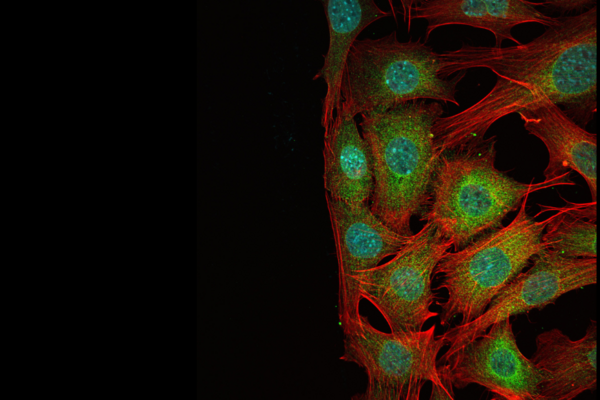
Second place 'The Wound Edge'
Ying (Angela) He, Undergraduate student
About Angela's image
Angela captured this image while completing research in the lab of Dr. Michelle Bendeck as part of her undergraduate studies in LMP. This is an immunostaining of MOVAS cells (mouse aortic smooth muscle cells) that have been injured by a scratch assay, hence the wound-edge where no cells are present. The image was taken using a laser-scanning confocal microscope (Olympus FV3000) at 60x objective lens. In the image, the cyan stained for the nucleus, the green for GSK3-beta and the red for filamentous actin.
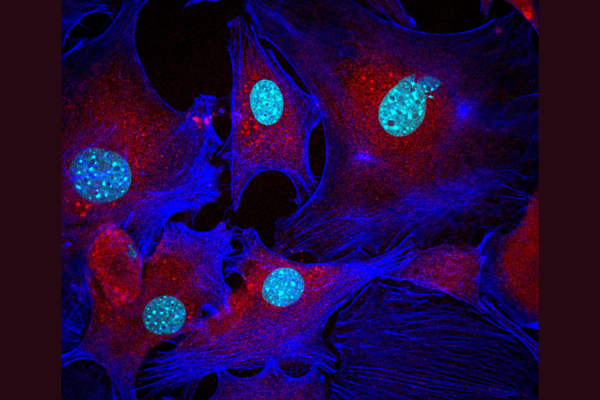
Third place 'Spiderman'
Ayla Shahid and Amanda Mao, MSc candidates in Dr. Michelle Bendeck's lab
About Ayla and Amanda's image
Vascular smooth muscle cells isolated from mice, stained for Yap (red) actin (blue) and nuclei (cyan). This image was taken with Olympus FV3000 confocal microscope at 60X magnification.
All submissions
It was a very difficult decision to choose a winner which you will see from all the wonderful entries below. Thank you to all who submitted an image.
Enjoy the beauty of LMP slides!
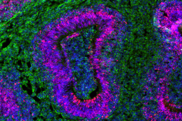
Rifat Sajid, PhD candidate
'I've Got Barney On My Mind': Immunofluorescence image of a brain organoid section highlighting neural stem cell-containing ventricle structures. SOX2, a neural stem cell marker is stained in red, while Beta III Tubulin, a neuron-specific cytoskeletal protein is stained in green. Cell nuclei are stained in blue. The overlap between blue and red in the ventricles produces the beautiful purple colour seen in the image, reminiscent of everyone's favourite dinosaur Barney.
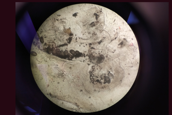
Greg German, Faculty
'A Rather Itchy Moon or Microscope: The Crusted Scabies Family': The optical microscope image (40x) of a skin scraping from a patient found to have crusted scabies. Typically scabies only has a few mites on the body at one time but those with a depressed immune system develop a crust or matte of scabies mites and eggs with thousands of mites. It is highly transmissible. In this picture, three adult mites with multiple eggs, feces, and larval stage mites.
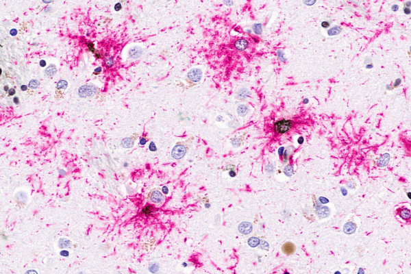
Seojin Lee, PhD candidate
'Fireworks of Tau': This is a brightfield microscopy image showing pathological tau proteins in pink and iron build-up in dark brown in the human brain with Progressive Supranuclear Palsy (PSP). Here we visualize cellular iron deposits in tau-positive tufted astrocytes, which are pathological hallmark of PSP.
Farbod Khorrami, MSc candidate
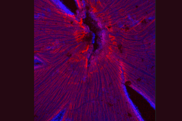
'Retinavigate: Highways of mouse vision': An immunofluorescence image of a whole-mounted mouse retina labeled with DAPI to label nuclei (Blue) and neurofilament to label axons (red). Axon bundles can be see radiating outward from the optic disc in the centre.
Ruchika Bagga, Resident
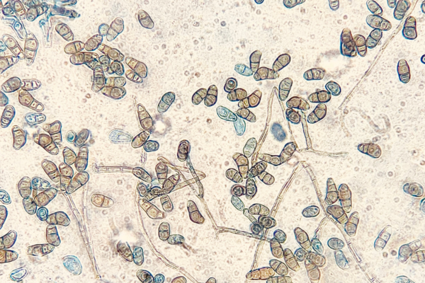
'No it’s not a Croissant! It’s Curvularia': A fun but rare fungal pathogen with croissant shaped conidia called Curvularia. Produces a black pigment hence called “ Dematiceous fungus”Image is under an Lactophenol mount under compound microscope.
Ain Kim, PhD candidate
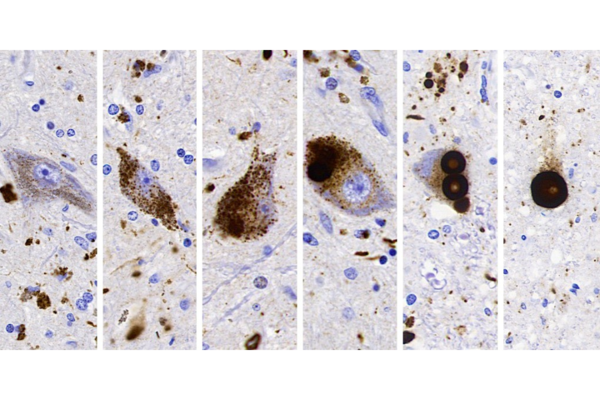
'Life of a Lewy Body': Lewy bodies in the substantia nigra of a Parkinson’s disease patient, immuno-stained with the 5G4 antibody that labels only the disease-associated α-synuclein.
From left to right, 1) a normal nerve cell with neuromelanin, a diseased nerve cell with: 2) pre-aggregates of α-synuclein, 3) small inclusions, and 4) a Lewy body. In rare cases, a nerve cell can be seen with 3 Lewy bodies! After a long journey, the life of a nerve cell comes to an end, leaving the Lewy body it formed.
Kayshani Kanagarajah, MSc candidate
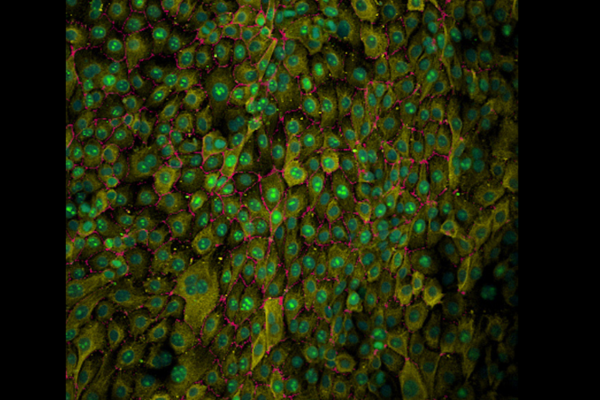
'Radiant Immortality': Immortalized human bronchial epithelial cells cultured on a dish and stained for Ki67 (green), ZO-1 (magenta), panCK (yellow), and nuclei (cyan). Imaged using Nikon A1 scanning confocal microscope.
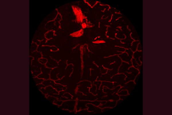
Arya Zarrinbakhsh, MSc candidate
'Overshot': A composite confocal microscope image depicting the choroidal vasculature of a mouse eye stained with isolectin B4. This tissue section was obtained after fortuitously overshooting coronal sections of the optic nerve and entering the choroidal space – a beautiful mistake.
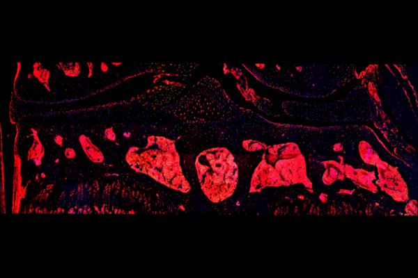
Byron Chan, PhD candidate
'Cell death in the knee': Fluorescent image of apoptotic cells along with their actin cytoskeleton in the mouse knee joint following Immunofluorescent staining with TUNEL (green), phalloidin (red), and DAPI (blue). Cross section of the mouse knee joint was imaged using a spinning disk confocal microscope. Images were stitched to capture the entire knee joint cavity.
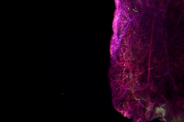
Nathaniel Vo, MSc candidate
'The Microcosmic Architecture of Adipose Tissue': This 3D image, captured using a light-sheet microscope, offers a glimpse into the interweaving network of vasculature (red) and sympathetic nerves (blue) that threads through the adipose tissue alongside the green hues of interspersing macrophages.
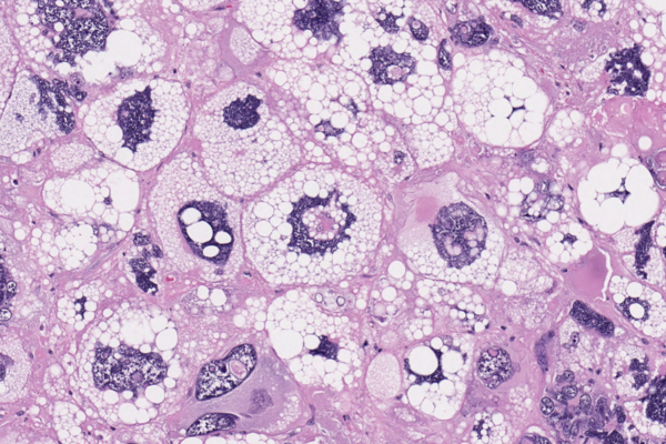
Shahd AlMohsen, Fellow
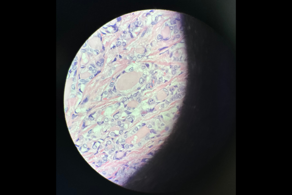
'Nucleus intra nucleum': This is a photomicrograph (H&E, x200) demonstrating a pleomorphic liposarcoma. The tumour is characterized by a spectacularly large number of lipid-laden cytoplasmic vacuoles, and so-called 'nucleus intra nucleum' (literally "nucleus inside nucleus").
Qianghua Zhou, Resident
'Unveiling the Crescent': Optical microscope. A Hematoxylin and Eosin Stain Reveals the Enigmatic Half Moon Pattern of Papillary Thyroid Carcinoma.
Mario Capitano, Faculty
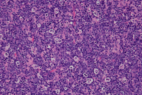
'Starry Night': This is an H&E image of a high grade lymphoma showing the 'starry sky pattern'. The night sky is composed of the lymphoma cells and the stars are composed of tingible body macrophages phagocytosing the nuclear debris of the rapidly dividing cells.
Ryunosuke Hoshi, PhD candidate
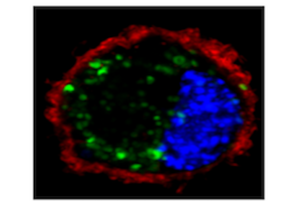
'Cancer on fire!': An immunofluorescence image of a murine lung carcinoma cell (CMT167) taken on the Quorum spinning disk confocal microscope using a 63x oil objective lens. Cells were fixed and stained with DAPI (blue, nucleus) and WGA (red, membrane) following delivery of fluorescently-labeled RNA (green) using ultrasound-mediated microbubble destruction (USMD).
Lusia Sepiashvili, Faculty
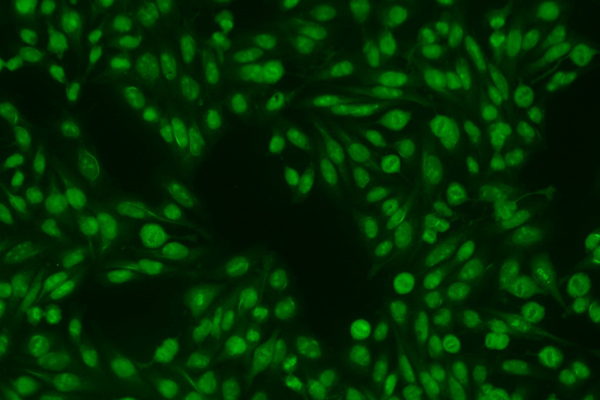
'Fluorescence': Indirect immunofluorescence of HEp-2 cells. A blood-based test used for evaluation of systemic autoimmune rheumatic diseases.
