Breadcrumbs
- Home
- About
- LMP Art Competition
- 2024 submissions & winners
LMP Art Competition winners 2024
Every spring, we ask the LMP community to show us the beauty of their speciality in the LMP Art Competition.
Our students, learners, faculty, and staff sent in many beautiful images and all members of the LMP community were invited to vote for their favourites online.
The top two images in each category receive a cash prize and a canvas of their image which will be displayed at the Annual Celebration of Excellence on June 13, 2024.
Enjoy the winners, and all submissions, of our art competition!
See submissions and winners from:
The following received the top votes from the LMP Community.
First place winners
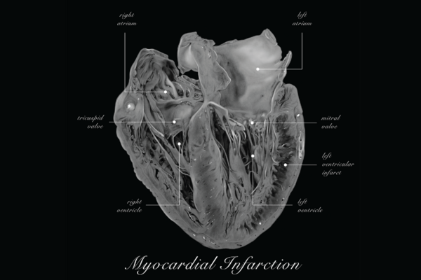
1st place: Clinical Laboratories
Cassie Hillock-Watling, MHSc Laboratory Medicine program (Pathologists' Assistant Field)
"Myocardial Infarction"
About Cassie's image
This is a pencil drawing of a cross section of a heart showcasing a myocardial infarction. After it was rendered on paper, it was edited using Procreate, Photoshop and Illustrator to add highlights, contrast and labels.
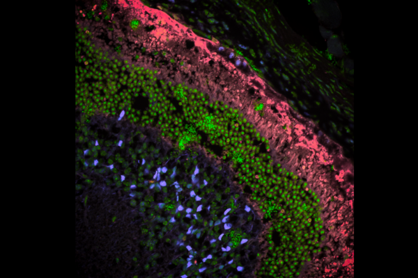
1st place: Basic Science Research Laboratories
Alissa Pak, PhD candidate in the Schuurmans Lab
"Flowers in the eyes"
About Alissa's image
The image portrays the retinal layers of the eye, metaphorically depicted as a garden. The Müller glial cells, highlighted with a nuclear marker Sox9 (purple), symbolize the seeds harboring the potential for regeneration. From these seeds, green sprouts emerge, representing the regenerative capacity of Müller glia to produce photoreceptors. These photoreceptors are depicted as the green nuclei (DAPI) and coral rod photoreceptors (Rhodopsin) symbolizing blossoming flowers. This analogy captures the dynamic nature of retinal cells and their pivotal role in maintaining vision.
Second place winners
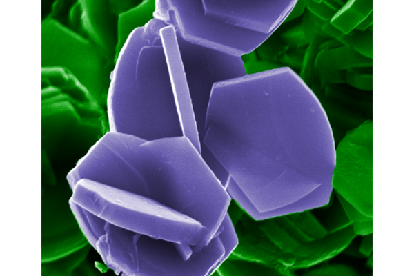
2nd place: Clinical Laboratories
Dr. Gino Somers, faculty member based at SickKids
"Sculpture on a nano scale"
About Gino's image
Colorized image of a urinary bladder stone, taken on a scanning electron microscope at a magnification of 5000 showing calcium oxalate crystals.
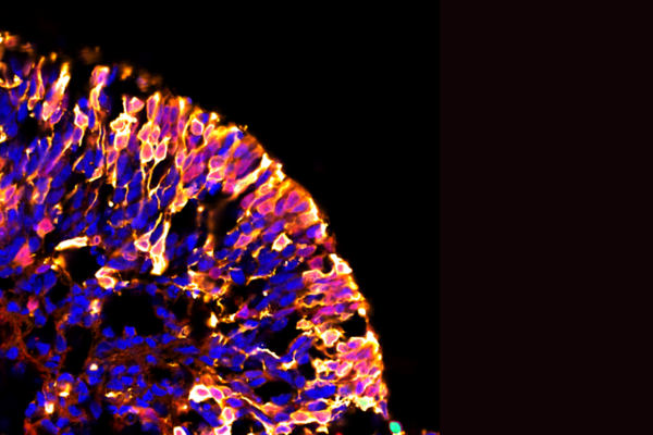
2nd place: Basic Science Research Laboratories
Jiajie Zhang, MSc student in the Ballios Lab
"Photoreceptors in retinal organoid"
About Jiajie's image
The light sensitive photoreceptors in our retina are the reason we have vision, they detect light that comes into our eye and transmit the signal into our brain to generate a visual image. This image is a close up look at a human stem cell-derived retinal organoid ("little 3D human retina grown in a dish"), with the photoreceptor cells (orange and red) on the outer edge of the organoids. The RECOVERIN staining (orange) reveals the photoreceptor cell morphology in the retinal organoids. This image is taken with confocal microscopy.
All submissions
It was a very difficult decision to choose a winner which you will see from all the wonderful entries below. Thank you to all who submitted an image.
Enjoy the beauty of LMP!
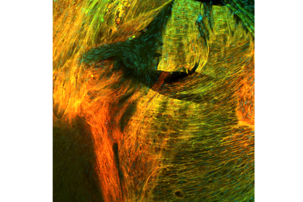
Ryan Appings, MSc student
Clinical category - "Precise delivery"
Laser scanning confocal microscope z-stack of N-cadherin mimetic peptide coated on nanoparticles which were delivered in vivo to an injured rat common carotid artery. Cyan (DAPI), Green (Elastin), Red (tetramethylrhodamine tagged peptide). The image depicts the superficial immediate elastin layer with fenestrations and the underlying medial layer with smooth muscle cells is exposed due to a tear.
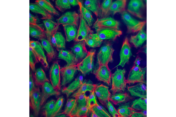
Shanti Mehta, Alumni
Basic Science category - "Cornea-copia"
Immunofluorescence microscopy of filamentous-actin (red), alpha-tubulin (green) and nuclei (blue) on the corneal endothelial layer (innermost layer of the cornea) of a patient with Fuchs endothelial corneal dystrophy.
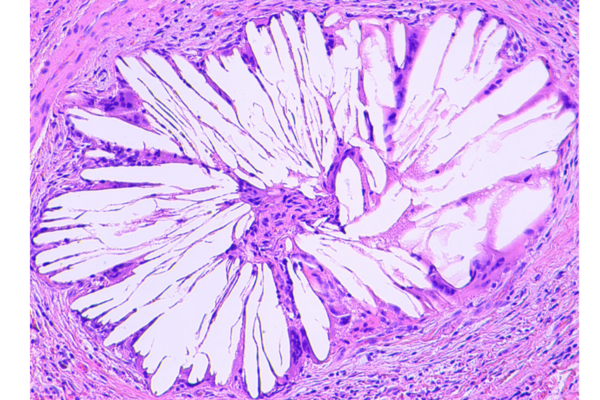
Monika Tripathi, Faculty member
Clinical category - "Mystic Moonflower"
"Hematoxylin & eosin-stained light microscopic image of Cholesterol granuloma within the gallbladder wall mimicking a rare moonflower species - Strophocactus wittii. The empty white spaces / flower petals are lipid and cholesterol crystals with pink multinucleate foreign body type giant cells around the edges.
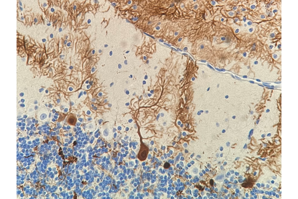
Lauren Joe, MSc Student
Basic Science category - "The Brain Forest"
DAB staining of a mouse brain injected with an adeno-associated virus (AAV) that expresses eGFP-tagged S3A cofilin (a constitutively active form of cofilin). Cofilin is an actin-binding protein that affects actin dynamics in ALS. Here we can see the branches of purkinje neurons in the cerebellum. This image was captured using brightfield microscopy.
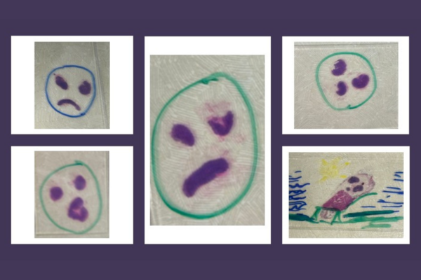
Dhuha Al-Sajee, Clinical Fellow
Clinical category - "Lymphemojis"
The image is a collection of approximately 15 lymph nodes on 5 slides, three in each. The last one is with salivary gland tissue. After a long work day and screening many lymph nodes for mets, you have to be creative and think outside the box to keep going. These lymph nodes were put in a way that reminded me of emojis.
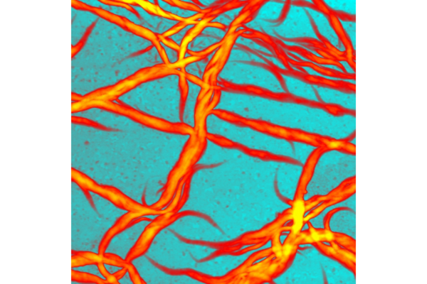
Laurent Bozec, Faculty member
Basic Science category - "Collagen as the threads of norms"
Collagen fibrils undergoing enzymatic digestion imaged by Atomic Force Microscopy (image size is 5x5 um).
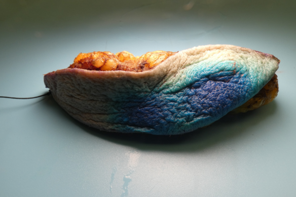
Michelle Malig, Pathologists' Assistant, UHN
Clinical category - "Skin in the game"
Gross macroscopic image of an oriented skin ellipse removed for Merkel Cell Carcinoma, stained with radioactive dye.
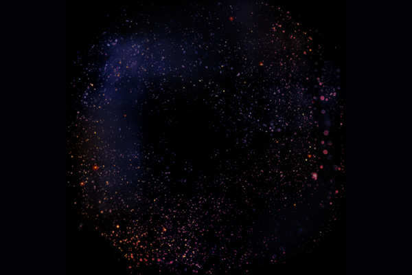
Ness Little, MSc student
Basic Science category - "Messier 64"
Reminiscent of outer space, Messier 64 is inspired by the ‘Black Eye Galaxy’ of the same title. The galaxy shows an immunofluorescence image of a whole human corneal endothelium transfected with adeno-associated virus (AAV). GFP expression is used to visualize cells infected with AAV2/5 and is shown here in purple and orange.
Ngoc-Nhu Jennifer Nguyen, Resident
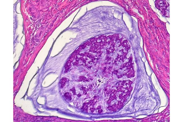
Clinical category - "Onigiri"
Bladder mass showing on light microscopy a mucinous adenocarcinoma. Haematoxylin & eosin.
Tobi Lam, Translational Research Program student
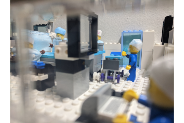
Basic Science category - "I forget your name but I know we work together"
This image depicts a model of the operating room, highlighting the people who are working in teams under dynamic uncertain conditions sometimes without knowing each other's names. I made this Lego model during the pandemic when we couldn't conduct research in-person.
Ramzi Fattouh, Faculty member
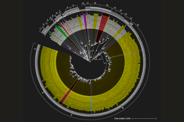
Clinical category - "Emergence and Spread of the SARS-CoV-2 C29200T Mutation in Canada"
Maximum-likelihood tree of SARS-CoV-2 in Canada (2020-2021) showcasing the emergence and spread of a mutation (C29200T) that caused gene-target dropout in a commonly employed diagnostic PCR test. Colours (yellow, orange, red, etc) indicate the province where the patient sample was collected. Uncoloured equals sequences without the mutation, used as controls. Sequence lineages also indicated. Colour/Image enhancements applied for display purposes.
Alena Zelinka, PhD candidate
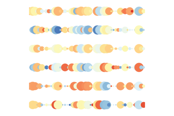
Basic Science category - "Diversity during development"
Gene expression fingerprints of cell populations in a developing cartilage organoid model, determined by single cell RNA sequencing. Circle colour and size represent average gene expression and percent of cells which express the gene of interest, respectively. Image created using RStudio and Seurat R package.
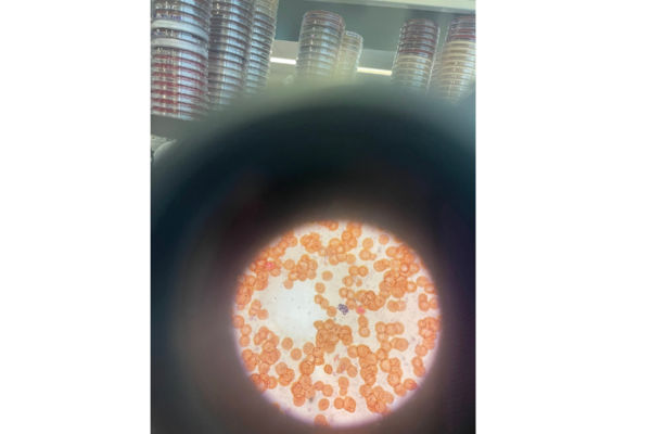
Gregory German, Faculty member
Clinical category - "Get Real (Sick) or Got Junk: at hospital blood culture bench"
View of a blood culture slide at 1000X final magnification containing gram-positive material in the centre, likely an artifact, with red blood cells and two lymphocytes present. In the background is a wall of used petri plates of different colours and ingredients used for culturing organisms. Blood cultures are the single most important test conducted in microbiology and require excellent collection technique to maintain quality and good patient care. While the test is over 100 years old there is a great deal of importance on reducing stain artifacts.
